Day 1
It’s the morning of our first day in Candiolo. I’m going there with Rosita and Debora : we are all very excited, even though we are still a little asleep. I’m also a bit scared about making any kind of mistakes , but I’ll try to do my best throughout these 3 days.
We left Cuneo early in the morning, and started our “alternanza” travelling to the research center. Once we got there, and trust me, we could tell a whole story just about how we tried to get there and come back every day , we asked the receptionist to help us find our tutor. He had no idea about who we were supposed to meet, so after he figured that out, he told us to go to the Molecular Oncology Dept.
When we finally arrived in the right department, we met our tutor Francesco. Well his name is Francesco but we actually called him Massimo for the whole day, we don’t even know why: we found out the mistake on the following day.
Anyway, Francesco showed us what he usually does when doctors take in the department an explanted tumor. He analyzes the genomic DNA : he tries to obtain only DNA out of cancer cells. Once he gets a filamentof DNA he puts it into a special machine which shows the different mutations affecting those cells. In Candiolo is paid a particular attention on the mutations affecting MET. Francesco taught us that the hardet part of the whole process is separating DNA from the other strucures of the cell. The tumor is cut in very light pieces and then the researcher adds different buffers, chemical solutions which destroy the bonds between the cells and the membranes, so that every single component of the cell is detached from the others. Then the mixture is heated up first at 37 °C for 30 ‘ and then at 50° for 2 hours. At the end of this, all we can find on the bottom of the falcon, once all the rests of the membranes have been absorbed with a liquid solution, is the DNA. At the end the filament is processed in a machine that uses ultraviolet rays to read it. The machine prints a chart where there are the results of the analysis on the DNA: the mutations affecting the different genes are highlighted. This time our experiment was looking for the mutations affecting only 3 genes that are usually mutated when cancer occurs (especially colon cancer). What surprised me is that the machine doesn’t only say if there’s a mutation., but it also recognizes how the mutation changes the basis. This is identified by the name of the protein produced instead of the right one: the proteins are the product of the expression of each gene, so mutations affect the whole production of proteins , which can be over produced or can be completely absent.
This part of the research in Candiolo is important because it says what’s really wrong in the DNA of a subject affected by cancer. Obviously this is not meant to test a particular cure, but to understand which genes are connected with different types of cancer.
Davide explained to us the difference between a benignant tumor and a malignant tumor. It basically consists in the ability that cancer has to infiltrate the tissues around the neoplasy, and, of course, the ability of producing metastasis. A tumor begins with a mutation in a single cell which then, replicating itself, creates a whole group of mutated cells. These cells then can have other different mutations, this is why usually tumors are eterogeneus: they are made of cells with different mutations. If a tumor is diagnosed in a really short time there might be a better chance to eliminate it, because of its little dimensions and because it wouldn’t be too eterogeneus yet. It’s important to notice that not all the mutations in a cell must be associated with cancer: just some of them in fact cause cancer! Many mutations are asymptomatic and are not the cause of any illnessness.
Nowadays the therapy against cancer is chemiotherapy , associated with radiotherapy. A patient has to be set in a cycle of these therapies even though the whole tumor has been removed because there might be metastasis that can’t be seen by any test like the PET or the TAC. Davive told us that in a few research centers in Italy scientists are studying a new machine, called TAC-PET, which combines the high resolution of the TAC with the capacity of PET of using a marker. In this way it would be possible to see very small marked metastasis in the body of the patient and prevent him from a possibly useless cycle of therapy.
Davide then took us to see how his project was going on. He wanted to see if some cells he had infected with artificial viruses were really infected. He had made artifcial viruses whose DNA coded for a special MET protein. This artificial version of MET, a membrane receptor, was able to be swiched on at taste. The virus’ DNA was marked with GFP, a luminescent protein. To be clearer, he wanted to see which of all the cells had absorbed the artificial DNA with that marker. The machine he wanted to use is called CELL SORTER.
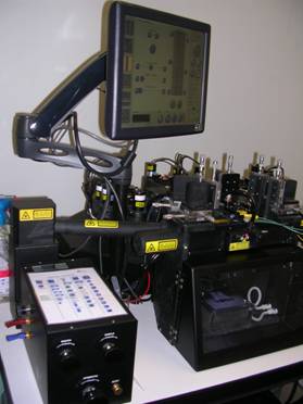
The sorter doesn’t only recognize the cells with the marker, but separates them from the ones that are not infected. This machine is really useful because it can divide different cells according to the marker which characterizes them in different test tubes. The result is a homogeneus population of cells. Davide explained to us how long it takes to get results from any kind of research. For example he has been working on his project for almost two years and he will have to leave it to another scientist once he is be done with his doctorate. He also showed us the importance of cooperation between doctors and scientists. These two worlds should be working to reach the same goal: the pursuit of better conditions for people, however sometimes fame and power get over the initial aim.
The first day was really interesting because we understood immediately what life in a researh center is like, which are the problems and which are the good things. It’s more or less like in the rest of the world: some people are trying to get personal success, while some other are trying to help people work real hard and sacrificing a lot of their personal needs.
Day 2
As soon as we got in Candiolo again, Davide wanted to show us what he usually does three times a week, every week. He has to replant the cells that he keeps in colture for all the experiments. He said that he used to work with many different populations, and he had to replace about 50 plates every time. Fortunately that day he had only four. This whole process is needed because cells replicate really fast and after a few days there are too many in a single plate, and they need more nourishment. Davide has to pay attention not to mix the different populations and to be careful when writing the right code on each plate. What’s hard to get is to separates the cells from the growth medium and the plate. Once this has been reached, he only replaces a fifth of the cells.
Davide showed us the different populations under the microscope , and it was really interesting to be able to see the different shapes of the cells and be able to understand their behaviour.
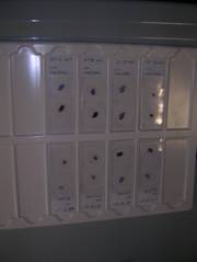
After that we met another researcher called Francesco, too. He is responsible for the immunohistochemistry department. In this part of the lab he basically does two things : he takes pieces of tumor as soon as they are explanted and puts them on slides so that they can be preserved forever, and he analyzes tissues marked with marked anticorps putting them on slides after putting a dye on them. For both the operations he first puts the piece of tumor in formaline and then is put in a block of paraffin. Then he uses a special machine called “microtomo” which cuts really thin slices of the frozen block of paraffin. Those slices are stuck on slides and from this point they can be dyed and then analyzed. This whole process is meant to preserve tumoral tissues and allows scientists to study how a tumor infiltrates tissues. Many of the tumors that Francesco has to work on are used to test the presence of antigens. Antigens are produced once the body recognize a viral attack or a change in its normal course of life, so finding a certain kind of antigens could tell the scientists what caused a change in the cell and the tumor. To find an antigen Francesco tests a slide adding an antibody. Knowing that antibodies stick only to the corrisponding antigen, he can, marking the antibody, see if it found its antigen.
This practice allows researches to understand which the factors that cause cancer are and gives the chance to see the effects on the tissues.
Francesco showed us an example testing a particular antibody on breast cancer cells already placed on a slide. Using this method a tumor connected with glands can be identified, so that it can be matched with a HT (hormone therapy).
Day 3
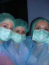
In my opinion this was the most interesting day because we saw something that we cannot see anywhere else: we visited the enclosure where the mice are kept. This is a separate area from the main building because of security for both the patients and the mice.To get inside it it’s necessary to wear albs, cuffs and gloves, and to walk through a special door that cleans the air so that people can’t bring microbes inside. The reason why it’s so dangerous to infect mice is that many of them are immunocompromised and couldn’t surive an infection. There are two separate rooms, in the first one there are the mice used for breeding and in the other one the mice that have a tumor implanted.The tumors are implanted between the skin and the muscle so that its growth doesn’t hurt the mouse,and it’s kept growing till it reaches 2000mm cubes of volume. When it happens the mouse is sacrified and the tumor is explanted. Usually tumors coming from a human patient are implanted, and right now in the center the research is concentrated over liver cancer and colon cancer. There are usually five mice per cage and they have food and water all the time. In the water there are usually anti-inflammatories so that mice can’t feel any pain. They are treated with particular drugs twice a day. In this way researchers can test different medicines on different tumors. Davide showed us how to give the drug to the mice, with a tiny tube pushed down into the stomach so that they cannot spit it out.They don’t feel any pain from this practice, but the scientist must be really good at doing it because a wrong movemnt could kill the mouse. He showed us different mice: imunocompromised ones without fur, black ones , some that were just born , some with a huge tumor and some with a little one just implanted. I wish it could be possible not to use animals to test medicines or anything else, but sometimes it is the only way to understand what the reaction of the whole body to a certain substance can be. I hope in the future we’ll find a better solution , but for now this is the only way to test something before testing it on a human being.
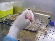
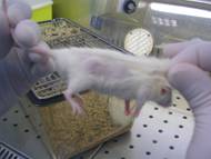
After that Davide showed us a tumor explant. He first anesthetized the mouse, and then, when it was completely asleep, he killed it with the tecnique of dislocation: using twisers to hold the neck, he pulled the tail till he heard the spinal cord break. At this point he cut the skin and explanted the tumor, dividing it into three parts: one for the genomic analysis, one for the immunohistochemistry and the last one for the quickfreezing, another way to preserve the tumor.
We spent the rest of the day next to the Cell sorter machine with Stefania, a researcher who was explaining her project to us. She was trying to separate the different cells that make up a breast cancer with different markers, using the sorter. She wanted to be able to state which are exactly the cells where the mutation occurs and the ones that are not affected at all. After this, she wanted to separate the cells again using the sorter to get homogeneus populatins of cancer cells. If she could separate these populations she could test these different drugs and the chances to find one that might actually work would be really high. The problem is that usually the sorter makes mistakes during the division, or there are just a few cells still alive. This is the reason why this is such a slow process: we spent a couple of hours with her and we had to go back to the laboratory twice to fix a problem with the colony of cells.
I am really satisfied with this experience, because it helped me to understand a world, the researh world, which is usually misunderstood or just left by. People don’t realize how hard it is to get a result out of a whole research, and how stressful it is to start everything all over again when something goes wrong. I strongly suggest that other students do this experience because it helped me decide what I want to do in my life, what I would like to become and how I want to be.
For example Davide taught me that collaboration is indispensable and that people should always keep in mind what the right goal to follow is. He is a great example for me because he is a doctor and he is also working in research. I think that those who do research should always be related with patients to be motivated and to be pushed to pursue the right aim.
In my opinion it is also important to inform people about what research really is and sensibilize them about this topic. A lot of people just think that a certain thing is wrong just because they were told that in the past, people should keep up with the research progress and understand how useful it is. In Italy there’s such a closed mentality about innovations and technology, that something is considered wrong and dangerous just because unknown!!
I hope in the future we, the young generations, will be able to help people understand the truth about the research world and that we will be supported and helped in every scientific field. I believe that if schools offered this kind of experiences the future could be really different. But for now, I hope I’ll be able to spread these pieces of information to the people around me at least. Someone once said that’s one small step for a man, a giant leap for mankind… we are responsible for our future, so we have to start to work hard to make it better. Every little thing we do can improve the situation a little, it can be about research, or medicine, or engeneering, or economy, or whatever, but we have to do somehing.
Well, now, after this experience, I’m much more convinced that research and medicine is what I’m interested in, those fields are the ones that I wish I could improve. I will work hard to make a change.
Chiara Paganelli – 4ª E – Anno scolastico 2009/10